
Файл:Development of the neural tube.png
Не е налична версия с по-висока разделителна способност.
Development_of_the_neural_tube.png (598 × 368 пиксела, големина на файла: 36 КБ, MIME-тип: image/png)
| Този файл е от Общомедия и може да се използва от други проекти.
Следва информация за файла, достъпна през оригиналната му описателна страница. |
Резюме
| Описание |
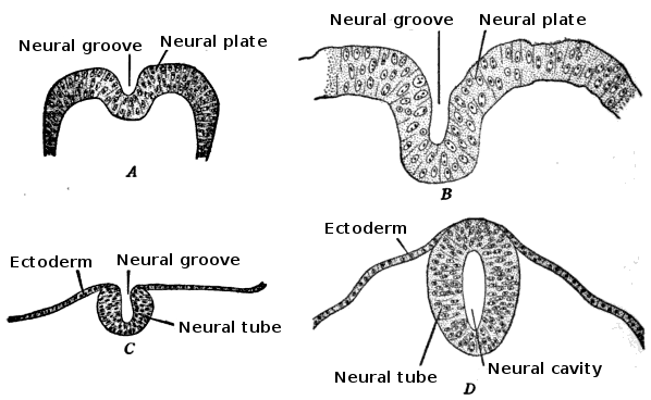
English: Development of the neural tube in human embryos (Prentiss-Arey). A. An early embryo (Keibel) B. at 2 mm. (Graf Spee) C. at 2 mm. (Mall) D. at 2.7 mm (Kollmann).
This is a scan of Figure 6 of the book "The anatomy of the nervous system" by Stephen Walter Ranson, with the labels redrawn. |
||||||||||||||||
| Дата | file created 2009-12-24, original image published 1920 | ||||||||||||||||
| Източник |
Figure 6 (p. 24) of "The anatomy of the nervous system" by Stephen Walter Ranson, published W.B. Saunders, 1920
|
||||||||||||||||
| Автор | user:Looie496 created file, original artist unknown | ||||||||||||||||
| други версии |
|
||||||||||||||||

== Лицензиране ==
This media file is in the public domain in the United States. This applies to U.S. works where the copyright has expired, often because its first publication occurred prior to January 1, 1929, and if not then due to lack of notice or renewal. See this page for further explanation.
|
 | |
 |
This image might not be in the public domain outside of the United States; this especially applies in the countries and areas that do not apply the rule of the shorter term for US works, such as Canada, Mainland China (not Hong Kong or Macao), Germany, Mexico, and Switzerland. The creator and year of publication are essential information and must be provided. See Wikipedia:Public domain and Wikipedia:Copyrights for more details.
|
История на файла
Избирането на дата/час ще покаже как е изглеждал файлът към онзи момент.
| Дата/Час | Миникартинка | Размер | Потребител | Коментар | |
|---|---|---|---|---|---|
| текуща | 20:08, 5 януари 2010 | | 598 × 368 (36 КБ) | Looie496 | {{Information |Description={{en|1=Development of the neural tube in human embryos (Prentiss-Arey). A. An early embryo (Keibel) B. at 2 mm. (Graf Spee) C. at 2 mm. (Mall) D. at 2.7 mm (Kollmann). This is a scan of Figure 6 of the book "The anatomy of |
Използване на файла
Следната страница използва следния файл:
Глобално използване на файл
Този файл се използва от следните други уикита:
- Употреба в af.wikipedia.org
- Употреба в ar.wikipedia.org
- Употреба в az.wikipedia.org
- Употреба в bs.wikipedia.org
- Употреба в en.wikipedia.org
- Употреба в fr.wikipedia.org
- Употреба в gl.wikipedia.org
- Употреба в hr.wikipedia.org
- Употреба в id.wikipedia.org
- Употреба в sr.wikipedia.org